![]()
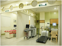
The SPH-T Cardiovascular Center performs standard and state-of-the-art diagnostic procedure to evaluate cardiovascular function and to identify structural abnormalities, including blockages or heart valve malformations.
It was established initially to provide specialized diagnostic services in Tuguegarao and the nearby town. It further developed to respond to the needs of the times thus the focus of using state-of-the-art technology, equipment and expertise was enhanced to ensure the most accurate diagnoses and treatment in a timely manner.
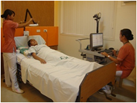
SPH-T's Cardiologists work together to provide patients with comprehensive cardiovascular services aimed at preventing and diagnosing cardiovascular diseases.
![]()
Services Offered:
A. Electrocardiography (ECG)
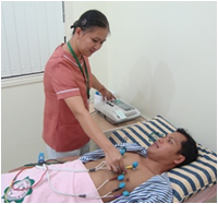
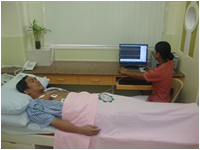
The E.C.G. is a on-invasive test that records the electrical activity of the heart through electrodes placed on the arms, legs and chest.
It provides information about heart rhythm and it also may giver evidence of whether or not a person has had a previous heart attack or is experiencing an on-going heart attack.
B. 24-Hour Holter Monitoring
The Holter Monitor is attached to the patient and is used to determine how the heart responds to normal activity.
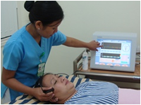
C. Electroencepalography (EEG)
The E.E.G. is a medical test used to measure the electrical activity of the brain via electrodes applied to the scalp.
It is used to detect abnormalities retaled to the electrical activity of the brain.
D. Transcranial Doppler Scan (TCD)
The (T.C.D.) is a medical test that ises a fast 3-5 seconds sweep speed that allows to see details of the waveform and spectrum.
It shortens the time necessary to find the window and to identify different arterial segments with a single gate spectral TCD.
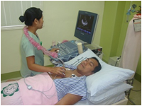
E. 2D- Echo
The 2D-Echo is a non-invasive test that uses sound waves to form a picture of the heart in motion.This allows evaluation of the function of the heart muscle and the heart valve.
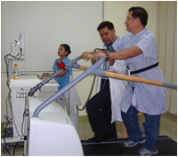
F. Stress Test/Treadmill Exercise
The Stress Test/ Treadmill Exercise is an exercise test used to evaluate how the heart responds to the stress of exercise.
This is accomplished by having the patient walk on a treadmill while the ECG is recorded.
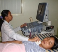
G. Peripheral Vascular Studies
The Peripheral Vascular Studies is an indirect, non-invasive ultrasound exam used to screen for peripheral vascular studies.
1. CAROTID ARTERY DOPPLER SCAN A Carotid Artery Doppler Scan is a type of Vascular Study done to assess the blood flow of the arteries that supply blood from the heart through the neck to the brain. It is also done to assess occlusion (blockage) or stenosis (narrowing) of the carotid arteries of the neck and/or the branches of the carotid artery.
2. PERIPHERAL VENOUS DOPPLER SCAN A Venous Ultrsound provides picture of the veins through the body that carry blood back to the heart. It can detect blood clots in the deep veins of the leg that can cause leg pain and swelling and can increase a person's risk of pulmonary embolism. It also evaluates abnormal veins causing varicose veins or other problem.
3. PERIPHERAL ARTERIAL DOPPLER SCAN


 through CHRIST-CENTERED Health Care Services
 Proclaim God's love to all...
 Value LIFE...
 Deliver holistic quality health care...
 Provide opportunities for total development...
 Pursue excellence and value innovations...
 The HEART of Compassionate Caring...